The Journal of Prosthetic Dentistry · 2025
What is the facial approach in graftless complete-arch rehabilitation?
In a clinical report published in The Journal of Prosthetic Dentistry (2025), Dr. Fernando Giovanella describes a way to place the anterior implants in a maxilla with severe atrophy: tilted to emerge facially rather than palatally, to reduce the anterior cantilever and enhance bone anchorage. Two zygomatic implants provide posterior support.
Graftless complete arch rehabilitation using a facial approach for enhanced bone anchorage and prosthetic outcomes: A clinical report
Fernando Giovanella
The Journal of Prosthetic Dentistry. 2025;134(2):295.e1-295.e6.
Abstract
Abstract of the accepted manuscript
This clinical report introduces a novel approach for the placement of anterior implants in maxillae with severe atrophy, with the objective of reducing anterior cantilever length and enhancing bone anchorage. By sagittally tilting the anterior implants to emerge facially rather than palatally, the procedure enhances prosthetic screw position and decreases the risk of a bulky prosthesis. Two zygomatic implants were placed posteriorly for additional support, and bilateral paranasal prostheses helped restore midfacial contours. Early follow-up showed stable implants and good soft tissue health. Longer-term studies and clear patient selection criteria are needed to confirm the effectiveness of the technique.
Introduction
Full text of the accepted manuscript
Graftless treatment options for complete arch rehabilitation have been shown to be predictable and effective,1–4 as an alternative to more invasive approaches.5,6 Posterior support has been successfully addressed through the tilting of posterior implants,7–13 the placement of pterygoid implants,14 or the use of zygomatic anchorage,3,15–20 effectively reducing or eliminating unfavorable posterior cantilevers.
Anterior anchorage in the maxillae has traditionally been achieved by utilizing the residual bone through a standard implant placed via a palatal approach21 or by employing zygomatic implants.14,15,19 More recently, transnasal implants, though still under development, have also been explored.22,23 However, the complete arch rehabilitation are, in some clinical scenarios, associated with significant prosthetic drawbacks.24,25
Since maxillary resorption in the anterior region progresses from anterior to posterior,26 it is common to encounter a Class III or pseudo-Class III intermaxillary relationship.27,28 This tends to lead to an anterior cantilever can be associated with a prosthesis with increased bulk, leading to screw access holes being positioned too far posteriorly relative to the prosthetic teeth. This unfavorable positioning may adversely affect speech, complicate hygiene, and compromise biomechanics.24,25,29
Addressing the effects of facial aging is also essential for achieving comprehensive therapeutic outcomes in edentulous patients. A variety of techniques, materials, and adjunctive solutions have been documented, which can be strategically implemented to optimize treatment results.30–40 This clinical report describes a novel surgical approach aimed at achieving optimal prosthetic outcomes in complex situations, while secondarily enhancing facial esthetics through adjunctive treatment.
Clinical report
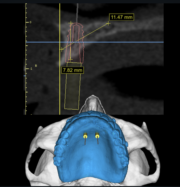
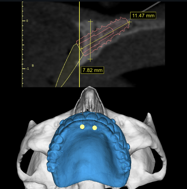
A 78-year-old female on levothyroxine and losartan presented with an atrophic maxilla. The lower arch had well-conditioned anterior teeth and implant-supported prostheses from the left first premolar to the left first molar and the right canine to the right first molar. A well-fitting maxillary denture, along with the well-aligned occlusal plane, ensured stable occlusion, serving as a reference for treatment planning (Fig. 1). Reverse treatment planning was performed using the digitized denture as a prosthetic reference in a software program (BlueSkyPlan; BlueSkyBio, LLC). Two 3.8×8-mm Morse taper conical-connection implants (EpikutCM; SIN Implant System) were simulated in the anterior region using a palatal approach. However, the evaluation revealed that the prosthetic screw emergence would be too far from the anterior dental axis (Fig. 2). To optimize the prosthetic outcome, the anterior implants were simulated in a transpalatal position, with the implant head emerging facially. This improved bone anchorage and allowed for a longer (3.8×11.5 mm) implant. A 30-degree angled mini-abutment enhanced the prosthetic screw’s emergence profile (Fig. 3). Two zygomatic implants were simulated for posterior support.
To evaluate the effect of omitting the flange in a fixed dentogingival prosthesis, a diagnostic trial prosthesis was created by digitally removing the flange from the left hemi-arch and 3D printing it in biocompatible resin (PriZma 3D Bio Prov; Makertechlabs). The evaluation revealed reduced dental exposure and a depression in the left labial region. Preoperative discussions confirmed the need for bilateral paranasal prostheses.
Under general anesthesia, a mucosa-supported surgical guide was employed for flapless initial demarcation of the osteotomy sites. Subsequently, a supracrestal incision was made along the maxillary ridge, complemented by 2 vertical incisions over the zygomatic buttress.
In the anterior region, the incision was made palatal to the initial implant perforations. A full-thickness mucoperiosteal flap was elevated, followed by osteotomies for placing 4 implants: 2 anterior implants and 2 posterior 4.0×40 mm zygomatic implants (ZygomaticPlus; SIN Implant System) (Fig. 4,5).
All implants achieved insertion torque values around 45 Ncm. Forty-five-degree angled abutments (Indexed 45-Angled Multi-Unit Abutment; SIN Implant System) were placed on the zygomatic implants and 30-degree angled abutments (Indexed 30-Angled Multi-Unit Abutment; SIN Implant System) were installed on from anterior implants. As planned, 2 paranasal prostheses were intraoperatively fabricated using polymethyl methacrylate (PMMA) surgical cement (C-MAXX; Cimtech), following a previously published protocol38 and secured with 2 titanium screws on each side in the paranasal area (Fig. 6). To enhance the anterior ridge volume and improve lip support, 0.5 g of hydroxyapatite (Osteogen; Intra-Lock) was applied.
The flap was closed using 4-0 polyglactin 910 sutures (Vicryl; Ethicon) (Fig. 7). Impressions were made, and a complete acrylic resin prosthesis with a metal infrastructure was delivered 3 days after surgery.
Recovery was uneventful. At 3.5 months, all implants had been successfully osseointegrated with healthy soft tissues (Fig. 8). A cone beam computed tomography (CBCT) scan confirmed accurate positioning of the bilateral zygomatic implants within the zygomatic bone and proper sagittal and facial emergence of anterior implants. The bilateral PMMA prostheses extended to the infraorbital nerve limits (Fig. 9) for enhanced facial esthetics (Fig. 10), and the definitive restoration showed optimal implant distribution, cantilever design, and tooth-screw distance (Fig. 11).
Figures 1 and 4 to 11 are clinical photographs and scans of the patient; they are in the accepted manuscript (PDF).
Discussion
The palatal approach, a well-established graftless technique for anterior implant placement within the All-on-4 concept, utilizes the residual bone for anchorage11,13; However, in cases like this, it may lead to excessive anterior cantilevering, bulky prostheses, and posteriorly positioned anterior screw access channels, potentially compromising speech, hygiene, and biomechanical stability.24,25,29 Furthermore, this approach fails to adequately address the challenges associated with lip support and the correction of deficiencies in the paranasal area.32
The combination of Le Fort I osteotomy and iliac bone graft effectively enhances facial esthetics and prosthetic outcomes in atrophic maxillae but requires extensive grafting.5,6 Using Le Fort I osteotomy with zygomatic implants offers a less graft-dependent alternative but is highly invasive and complex.41
Less invasive techniques, such as the utilization of particulate xenografts in conjunction with collagen membranes,19 or a pedicled fat pad,42 have been proposed to enhance the sagittal support of the superior lip. The primary effects of these interventions could improve the lip support, but do not adequately address the anterior cantilever challenges associated with the palatal positioning of the implant and the prosthetic outcomes.24,25 Similarly, challenges persist with transnasal implants and zygomatic anchorage.
The facial approach, as described in this clinical report, presents some potential advantages. By placing the implants in a more horizontally or tilted position, with the implant head emerging from the facial aspect of the residual ridge, the anterior cantilever length is minimized, leading to a more favorable prosthetic outcome. This technique allows the use of longer implants, thereby increasing bone anchorage. The utilization of angled abutments facilitates precise adjustment and optimal positioning relative to the definitive restoration. In the current patient, a 30-degree abutment provided adequate correction.
Despite the unconventional nature of this approach, the biomechanical principles governing implant dentistry remain consistent. Critical implant techniques that involve the placement of implants in a tilted fashion in posterior regions, subject to high occlusal forces,43 and the extension of some implants beyond the bone envelope, such as tilted transsinus or extramaxillary zygomatic implants, deviate from conventional intraosseous approaches. However, they still adhere to the principles of osseointegration and demonstrate a high success rate.3,10,12 The All-on-4 concept demonstrates a high success rate, even when the implant insertion torque is low.11
In the facial approach, the biomechanics are comparable to those of distally tilted implants, which are well-supported by evidence7–10; however, the implant remains entirely within the bone envelope and is tilted facially, positioning it in a region subjected to relatively lower occlusal forces compared to the molar area.43 In contrast to the zygomatic externalized technique,3 where implant threads are exposed in the buccal region, this novel approach ensures that no implant threads are exposed.
Conversely, the facial approach presents certain limitations. One key concern is the facial emergence of the implant head and abutment in an area lacking keratinized mucosa. Although keratinized mucosa is generally recommended44, maintaining optimal oral hygiene, as demonstrated in this case, may help prevent soft tissue complications. However, soft tissue grafting can be considered for future cases.
Facial aging involves a clockwise rotation of the maxillae, necessitating consideration of skeletal changes for effective treatment. In maxillary complete dentures, the labial flange is crucial for lip support and esthetics. However, in cases of severe maxillary atrophy, traditional flanges often fail to restore midfacial contours, particularly in the paranasal region.3–33
Paranasal prostheses address these deficiencies by supporting nasolabial and labial tissues, enhancing esthetics and patient satisfaction. Various materials can be used,34–37,40 but polymethyl methacrylate (PMMA) was chosen for its transsurgical molding capability and proven craniofacial safety.39
Summary
This clinical case demonstrates the possibility of a facial approach for anterior implant placement in graftless complete-arch rehabilitations, addressing the challenges of severe maxillary atrophy. By tilting the implant sagittally to emerge from the facial side of the maxillary ridge, this approach enhanced bone anchorage, reduced anterior cantilever, and improved prosthetic outcomes. Paranasal prostheses enhanced midfacial support and aesthetics. Further research is needed to validate the long-term efficacy of the facial approach and refine guidelines.
Patient consent
Written informed consent was obtained from the patient for the publication of this case report and accompanying images.
References
- Davó R, Fan S, Wang F, Wu Y. Long-term survival and complications of Quad Zygoma Protocol with Anatomy-Guided Approach in severely atrophic maxilla: A retrospective follow-up analysis of up to 17 years. Clin Implant Dent Relat Res. 2024 Apr;26:343–55.
- Mehta SP, Sutariya PV, Pathan MR, Upadhyay HH, Patel SR, Kantharia NDG. Clinical success between tilted and axial implants in edentulous maxilla: A systematic review and meta-analysis. J Indian Prosthodont Soc. 2021;21:217–28.
- Migliorança RM, Sotto-Maior BS, Senna PM, Francischone CE, Cury AADB. Immediate occlusal loading of extrasinus zygomatic implants: A prospective cohort study with a follow-up period of 8 years. Int J Oral Maxillofac Surg. 2012 Sep 1;41:1072–6.
- Tavelli C, Tedesco A. Survival and complication rate of zygomatic implants: a systematic review. J Oral Implantol. 2022;48:123–9.
- Pieri F, Lizio G, Bianchi A, Corinaldesi G, Marchetti C. Immediate Loading of Dental Implants Placed in Severely Resorbed Edentulous Maxillae Reconstructed With Le Fort I Osteotomy and Interpositional Bone Grafting. J Periodontol. 2012;83:963–72.
- Terheyden H. A new technique of Le Fort I interpositional grafting for dental implants. Int J Oral Maxillofac Surg. 2017;46:53–4.
- Malak AA, El Masri Y, El Masri J, Issawi HA, Salameh P, Aoun G. Implant Failure and Marginal Bone Loss Between Axial and Tilted Implants: An Umbrella Review with Meta-analysis. Int J Oral Maxillofac Implants. 2024;11;875–83.
- Cortés-Bretón Brinkmann J, García-Gil I, Pedregal P, Peláez J, Prados-Frutos JC, Suárez MJ. Long-Term Clinical Behavior and Complications of Intentionally Tilted Dental Implants Compared with Straight Implants Supporting Fixed Restorations: A Systematic Review and Meta-Analysis. Biology. 2021;10:509.
- Chrcanovic BR, Albrektsson T, Wennerberg A. Tilted versus axially placed dental implants: a meta-analysis. J Dent. 2015;43:149–70.
- Maló P, Nobre M de A, Lopes A. Immediate loading of ‘All-on-4’ maxillary prostheses using trans-sinus tilted implants without sinus bone grafting: a retrospective study reporting the 3-year outcome. Eur J Oral Implant. 2013;6:45–52
- Maló P, Lopes A, de Araújo Nobre M, Ferro A. Immediate function dental implants inserted with less than 30N·cm of torque in full-arch maxillary rehabilitations using the All-on-4 concept: retrospective study. Int J Oral Maxillofac Surg. 2018;47:1079–85.
- Aalam AA, Krivitsky-Aalam A, Zelig D, Oh S, Holtzclaw D, Kurtzman GM. Trans-sinus dental implants, for immediate placement when insufficient alveolar height is present: an alternative to zygomatic implants – surgical case series. Ann Med Surg. 2023;85:51.
- Soto-Penaloza D, Zaragozí-Alonso R, Penarrocha-Diago M, Penarrocha-Diago M. The all-on-four treatment concept: Systematic review. J Clin Exp Dent. 2017;9:e474–88.
- Araujo RZ, Santiago Júnior JF, Cardoso CL, Benites Condezo AF, Moreira Júnior R, Curi MM. Clinical outcomes of pterygoid implants: Systematic review and meta-analysis. J Cranio-Maxillofac Surg. 2019;47:651–60.
- Candel-Martí E, Carrillo-García C, Peñarrocha-Oltra D, Peñarrocha-Diago M. Rehabilitation of atrophic posterior maxilla with zygomatic implants: review. J Oral Implantol. 2012;38:653–7.
- Aparicio C, Manresa C, Francisco K, Aparicio A, Nunes J, Claros P, et al. Zygomatic Implants Placed Using the Zygomatic Anatomy-Guided Approach versus the Classical Technique: A Proposed System to Report Rhinosinusitis Diagnosis. Clin Implant Dent Relat Res. 2014;16:627–42.
- Lan K, Wang F, Huang W, Davó R, Wu Y. Quad Zygomatic Implants: A Systematic Review and Meta-analysis on Survival and Complications. Int J Oral Maxillofac Implants. 2021;36:21–9.
- Tuminelli FJ, Walter LR, Neugarten J, Bedrossian E. Immediate loading of zygomatic implants: A systematic review of implant survival, prosthesis survival and potential complications. Eur J Oral Implantol. 2017;10 Suppl 1:79–87.
- Hernández-Alfaro F, Ragucci GMM, Giralt-Hernando M, Caramês J, Valls-Ontañón A. ‘Pillow technique’ to improve lip support in the context of zygoma implant rehabilitation. Int J Oral Maxillofac Surg. 2023;52:716–21.
- Gracher AHP, de Moura MB, da Silva Peres P, Thomé G, Padovan LEM, Trojan LC. Full arch rehabilitation in patients with atrophic upper jaws with zygomatic implants: a systematic review. Int J Implant Dent. 2021;7:17.
- Lekholm U, Sennerby L, Roos J, Becker W. Soft tissue and marginal bone conditions at osseointegrated implants that have exposed threads: a 5-year retrospective study. Int J Oral Maxillofac Implants. 1996;11:599–604.
- Gelpi F, Alberti C, De Santis D, Bevilacqua M, Mellone F, Tealdo T. A Retrospective Multicentric Study of 52 Nasal and Trans-nasal Implants in 31 Severe Atrophic Patients to Reduce Anterior Cantilever Bending in Full Arch Implant-supported Fixed Rehabilitation. Int J Oral Maxillofac Implants. 2024;0:1–21.
- Almeida PHT, Cacciacane SH, Arcazas Junior A. Extra-long transnasal implants as alternative for Quad Zygoma: Case report. Ann Med Surg 2012. 2021;68:102635.
- Block MS. Maxillary Full Arch Restorations - Biological Complications: A Narrative Review Outlining Criteria for Long Term Success. J Oral Maxillofac Surg Off J Am Assoc Oral Maxillofac Surg. 2023;81:1124–34.
- Van Lierde KM, Browaeys H, Corthals P, Matthys C, Mussche P, Van Kerckhove E, et al. Impact of fixed implant prosthetics using the ‘all-on-four’ treatment concept on speech intelligibility, articulation and oromyofunctional behaviour. Int J Oral Maxillofac Surg. 2012;41:1550–7.
- Sutton DN, Lewis BRK, Patel M, Cawood JI. Changes in facial form relative to progressive atrophy of the edentulous jaws. Int J Oral Maxillofac Surg. 2004;33:676–82.
- Cawood JI, Howell RA. A classification of the edentulous jaws. Int J Oral Maxillofac Surg. 1988;17:232–6.
- Tan WL, Wong TLT, Wong MCM, Lang NP. A systematic review of post-extractional alveolar hard and soft tissue dimensional changes in humans. Clin Oral Implants Res. 2012;23 Suppl 5:1–21.
- Brunski JB. Biomechanical aspects of the optimal number of implants to carry a cross-arch full restoration. Eur J Oral Implant. 2014;6:45–50
- Pessa JE. An algorithm of facial aging: verification of Lambros’s theory by three-dimensional stereolithography, with reference to the pathogenesis of midfacial aging, scleral show, and the lateral suborbital trough deformity. Plast Reconstr Surg. 2000;106:479–88; discussion 489-490.
- Mendelson B, Wong CH. Changes in the Facial Skeleton With Aging: Implications and Clinical Applications in Facial Rejuvenation. Aesthetic Plast Surg. 2012;36:753–60.
- Rajali A, Mustafa NW@ NA, Zulkiflee MZA, Rahman ANA, Ahmad R. The Impact of The Labial Flange and Artificial Teeth on Lip Support in Elderly Patients with Maxillary Complete Dentures. J Dent Indones. 2022;29:127–33.
- Yuan F, Cheng C, Dai N, Sun Y. Prediction of aesthetic reconstruction effects in edentulous patients. Sci Rep. 2017;7:18077.
- Kim JH, Jung MS, Lee BH, Jeong HS, Suh IS, Ahn DK. Silicone Implant-Based Paranasal Augmentation for Mild Midface Concavity. Arch Craniofacial Surg. 2016;17:20–4.
- Zhang JY, Liu K, Liu RX, Xu BH. Safety and Efficacy of Midface Augmentation Using Bio-Oss Bone Powder and Bio-Gide Collagen Membrane in Asians. J Clin Med. 2023;12:959.
- Zhang J, Li D, Liu Y, Zhang Z, Fan Y, Shi J, et al. Application of Patient-Specific PEEK Implant for Aesthetic Considerations in Paranasal Augmentation. J Craniofac Surg. 2022;33:e877–80.
- Kwon TG, Kang SM, Hwang HD. Three-dimensional soft tissue change after paranasal augmentation with porous polyethylene. Int J Oral Maxillofac Surg. 2014;43:816–23.
- Esteves L, Rodrigues D, Ávila C, Campos P, Santos J. Preenchimentos estéticos na cirurgia ortognática: há indicações? Rev Clínica Ortod Dent Press. 2016;15:33–59.
- Leão RDS, Maior JRS, Lemos CADA, Vasconcelos BCDE, Montes MAJR, Pellizzer EP, et al. Complications with PMMA compared with other materials used in cranioplasty: a systematic review and meta-analysis. Braz Oral Res. 2018;32:1–9.
- Aubry S, Collart-Dutilleul PY, Renaud M, Batifol D, Montal S, Pourreyron L, et al. Benefit of Hyaluronic Acid to Treat Facial Aging in Completely Edentulous Patients. J Clin Med. 2022;11:5874.
- Campos O, Pintor F, Zivov A, Dallaserra M, Oyarzún C. Lefort I and zygomatic implant for severe atrophic maxilla: a literature review and technique description. J Oral Maxillofac Surg 2021;79:1–10.
- Rubio-Bueno P, Ardanza B, Piñas L, Murillo N. Pedicled Buccal Fat Pad Flap for Upper Lip Augmentation in Orthognathic Surgery Patients. J Oral Maxillofac Surg. 2013;71:e178–84.
- Kayumi S, Takayama Y, Yokoyama A, Ueda N. Effect of bite force in occlusal adjustment of dental implants on the distribution of occlusal pressure: comparison among three bite forces in occlusal adjustment. Int J Implant Dent. 2015;1:14.
- Mancini L, Strauss FJ, Lim HC, Tavelli L, Jung RE, Naenni N, et al. Impact of keratinized mucosa on implant-health related parameters: A 10-year prospective re-analysis study. Clin Implant Dent Relat Res. 2024;26:554–63.
About this version
This is the accepted manuscript: the author's text after peer review, before the journal's copyediting and typesetting. The version of record, the one to cite, is published in The Journal of Prosthetic Dentistry.
© 2025. This manuscript version is made available under the CC BY-NC-ND 4.0 license.
How to cite
Giovanella F. Graftless complete arch rehabilitation using a facial approach for enhanced bone anchorage and prosthetic outcomes: A clinical report. J Prosthet Dent. 2025;134(2):295.e1-295.e6. doi:10.1016/j.prosdent.2025.03.046